Menorrhagia & Easy Bruising
June 27, 2001
Each issue, Q Fever! presents a challenging clinical conundrum to test readers' problem-solving skills and illustrate bread-and-butter medical principles. Good luck!
You are asked to evaluate a 19 year old white female who presents to your office with the chief complaint of menorrhagia and easy bruising.
Since the onset of menses at age 13, she has used approximately 9 pads per day for seven days during each menstrual cycle. She has also noted the frequent development of large (>3 cm) ecchymoses due to minor trauma since early childhood, along with bleeding gums when brushing her teeth vigorously, and occasional nosebleeds.
Other than these issues, she has no medical problems, and has never had major surgery.
She takes no medications and has no known drug allergies; she does not smoke or drink alcohol, and is a sophomore in college, where she lives with a roomate.
Her family history is significant in that her mother also had multiple bleeding problems, and suffered from severe menorrhagia throughout adolescence as well. In addition, a maternal great aunt died of post-operative bleeding in the 1940's.
On physical exam, the patient is in no apparent distress, and is very pleasant.
Temperature is 97.7F, pulse is 80, blood pressure is 120/70, and respirations are 12.
There is a 3 cm ecchymosis on her left upper arm, as well as a smaller one on her right shin.
Head and neck, lung, heart, and abdominal exams are normal, as is a pelvic exam.
Screening laboratory studies show a moderate microcytic anemia. The remainder of the tests, including PT/PTT and platelet count, are reported to be normal.
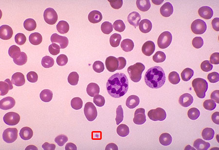
Hazily remembering something you learned during your medical school years, you visit the lab yourself and examine the patient's peripheral blood smear at 10x magnification:

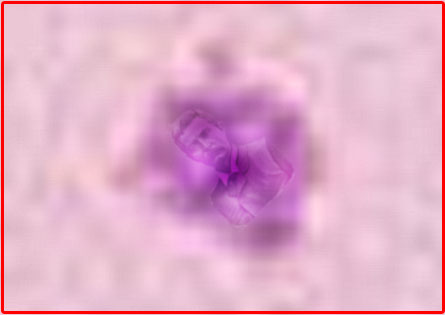
Viewing the outlined area at 500x magnification, you discover the following:

What's going on?
Answer:
Von Willebrand's Disease
This woman, who presented with menorrhagia and easy bruising, has von Willebrand's Disease.
The presence of Finnish physician Dr. Erik von Willebrand in the patient's platelets is demonstrated in the high-power view above, and is pathognomonic for the disorder.
Without a strong clinical suspicion, clinicians often neglect to utilize the 500x magnification lens necessary to visualize platelets containing Dr. von Willebrand with conventional light microscopy. Even then, von Willebrand is often mistaken for the Golgi apparatus, endoplasmic reticulum, or other typical cell structure, leading to an unnecessary delay in diagnosis.
It is important to remember that, while Dr. von Willebrand is most likely to be found in a classic frontal portrait pose, it is not unusual to see him lying comfortably on his side, or playing a round of golf with his friends.
The diagnosis can be confirmed by in vitro ristocetin challenge, which will cause von Willebrand's hair to become visibly stringy and dry, eventually falling out in clumps.
Treatment is straightforward and involves intravenous desmopressin (DDAVP), a substance which gently persuades Dr. von Willebrand to leave the affected platelets and return to his native village in the Scandanavian hinterland.
More Stuff!
Get the Q Fever! Book!
The Q Fever! Store!: T-shirts, caps, mugs, and thongs!
Support The Q!
Subscribe to the Q Fever! Mailing List!
Contact Q Fever!
Remember: Quality Without The Q Is Just Uality!
Menu

